Cells, Organelles and Mitosis
|
Objectives |
Learning Activities |
Resources |
|
1.1.1 Discuss the theory that living organisms are composed of cells. "Cell Theory" 1.1.3 State that all cells are formed from other cells. 1.1.11 Explain that cells in multicellular organisms differentiate to carry out specialized functions by expressing some of their genes but not others. 1.1.12 Define tissue, organ, organ system 1.1.4 Explain three advantages of using light microscopes. 1.1.5 Outline the advantages of using electron microscopes (the terms resolution magnification should be explained) 1.1.8 Calculate linear magnification of drawings |
Microscopes & Cells Revision of the use of light microscopes. Preparation of slides of tissues (including onion, celery skin, tradescantia leaves) using new stains. Make drawings of eukaryotic cells using light microscopes and make measurements using graticules. Question 2 p17 of Fullick on eukaryote cells. Understand measurement units, mm,mm,nm and how to calculate them & present them on drawings. Print six magnified images and on each one label either a magnification (e.g. x200) or a scale bar. TEST on MICROSCOPES |
Interactive Scanning Electron Microscope
|
|
1.1.6
Define organelle
1.3.1 Draw a diagram to show the ultrastructure of a generalized animal cell as seen in electron micrographs. 1.1.7 Compare the relative sizes of molecules, cell membrane thickness, viruses, bacteria,organelles and cells, using appropriate SI units 1.3.1 State one function of each of these organelles: ribosomes, rough endoplasmic reticulum lysosome, Golgi apparatus, mitochondrion, nucleus. 1.3.4 Describe three differences between plant and animal cells. 1.3.5 State the composition and function of the plant cell wall. |
Organelles Describe the function of the organelles found within these cells using electron micrographs. Diagram Using electron micrographs of organelles, learn to recognize the structure of; nucleus, chloroplast, mitochondrion, golgi apparatus, rER, Lysosomes, ribosomes. Presentation to the class on a single organelle's structure and function. Question 5 p86 Fullick on Prokaryotes and Lysosomes. |
More plant organelle electron miscroscope photos
|
|
1.2.1 Draw a generalized prokaryotic cell as seen in electron micrographs 1.2.2 State one function for each of the following: cell wall, plasma membrane, mesosome, cytoplasm, ribosomes, naked DNA. 1.2.3 State that prokaryotes show a wide range of metabolic activity including fermentation, photosynthesis and nitrogen fixation. 1.3.3 Compare prokaryotic and eukaryotic cells |
Prokaryotes View Bacteria on this link , and this one and describe the ultrastructure of prokaryotic cells and their organelles.
Experiment
to see gram stained bacteria. Grow some fluorescent Prokaryotes in the lab. |
The way antibiotics kill bacteria. Anthrax in particular. |
|
1.1.2 State that a virus is a non-cellular structure consisting of DNA or RNA surrounded by a protein coat. |
Viruses Are Viruses living? A look beyond the resolution of the light microscope. |
Viruses, their structure and why penicillin can never cure the flu? |
| EXAM TECHNIQUES |
Comparison style questions in IB Biology
TEST on CELLS |
Summary
IB Questions
|
|
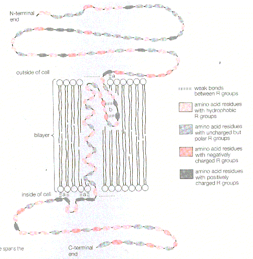
1.4.1
Draw a diagram to show the fluid mosaic model of a biological
membrane.
1.4.2 Explain how the hydrophobic and hydrophilic properties of phospholipids help to maintain the structure of cell membranes. 1.4.3 List the functions of membrane proteins1.4.3 Define diffusion & osmosis. 1.4.5 Explain passive transport across membranes in terms of diffusion. 1.4.6 Explain the role of protein pumps and ATP in active transport across membranes. 1.4.7 Explain how vesicles are used to transport materials within a cell between the rough endoplasmic reticulum, Golgi apparatus and plasma membrane. 1.4.8 Describe how the fluidity of the membrane allows it to change shape, break and reform during endocytosis and exocytosis. 1.1.10 State that unicellular organisms carry out all the functions of life. 1.1.9 Explain the importance of the surface area to volume ratio as a factor limiting cell size |
Membranes - the plasma membrane. The Fluid Mosaic Model of Cell Membrane; Learn the parts of the membrane using the Paint Exercise (file in Mr Faure's my photos) Study this diagram of membrane proteins and prepare a presentation on one of the above special functions of membranes. Observe diffusion in vitro. Complete practical investigation of osmosis using visking tubing and onion cells. Active and Passive transport. Protein pumps, diffusion chanels, vesicles, endocytosis, exocytosis. Solve the text book problem on function of a membrane protein Investigation of membrane permeability using beetroot. How big is an Amoeba? What is the largest cell. Why don't cells come bigger?
TEST on MEMBRANES |
Membrane part functions Powerpoint Activity
Animation of transport through a cell membrane (excellent)
|
|
1.5.1
State that the cell-division cycle involves interphase, mitosis and
cytokinesis.
1.5.2 State that interphase is an active period 1.5.3 Describe the events that occur in the four phases of mitosis1.5.4 Explain how mitosis produces two genetically identical nuclei. 1.5.5 Outline the differences in mitosis and cytokinesis between animal and plant cells. ie; the lack of the centrioles in plant cells and the formation of the cell plate. 1.5.6 State that growth, tissue repair and asexual reproduction involve mitosis 1.5.7 State that tumours (cancers) are the result of uncontrolled cell division and that these can occur in any organ. |
Mitosis - How it happens and its uses. Little java explanation Experiment
to make slides of root squashes to observe stages of mitosis.
The method is in the expt text book p184. Study the structure of DNA and chromosomes and the processes of transcription, DNA replication and the cell cycle. Detailed website on mitosis and cell cycle. FINAL Topic 1 TEST |
Prepare a power point presentation of the four stages of mitosis. Lab drawings of the stages I,P,M,A,T in plants and animals. Write a short informative leaflet explaining the biology of cell division and its role in Cancer. Learn about the cell cycle using this web site.
|
|
Extension Work Ideas |
Download a whole website of diagrams about cells. Try to do a Hot Potato Quiz. Lots of cell links
|
Bird Flu 2005 - will millions die? |