Bacterial Gram Stain Technique
After completing this laboratory, the students will be able to:
- Demonstrate sterile technique when preparing a bacterial smear for staining.
- Demonstrate proper gram staining procedures.
- Identify bacteria as being gram positive or gram negative.
Preparation;
Set up one work area with a Bunsen burner for heat fixing the bacteria.
Set up a staining area, away from the Bunsen burner, with the following items:
- Crystal Violet stain.
- Grams' stain.
- Safranin stain.
- Washing bottles with distilled water.
- 95% ethanol solution in a washing bottle.
- Slide holder
- Bacteria on smears.
All of these solutions stains can be in little dropper bottles labeled appropriately.
Use bacteria listed as safe for science classes.
When you have completed the lab, dispose of the slides in a sterile manner by putting them in a solution of 10% bleach. Wipe down the tables and the work areas with a 10% bleach solution.
Sterile Technique
Purpose of Sterile Technique:
- Protect You and the Environment from contamination by bacteria.
- Protect the Original Bacterial Culture from contamination.
1. Flaming: The wire can be sterilized by heating it over a bunsen burner until it is red hot. You start flaming the wire loop from the handle connection up to the tip. This will prevent the liquid at the tip from spraying into the air. Once the needle cools it can then be used to transfer bacteria.
2. Preventing Contamination of a Culture tube: Step by step procedure
- Pick up culture tube from rack with non-dominant hand and and wire loop with dominant hand.
- Flame wire loop.
- Remove cap from culture tube - hold cap.
- Flame culture tube lip.
- Insert loop end of wire loop into culture tube and remove a small sample.
- Flame lip of culture tube and replace cap.
- Put culture tube back into the rack from where it came.
- Streak sample over slide or ?
- Flame wire loop.
Gram Staining Procedure
Introduction:
 |
In 1884, Hans Christian Gram, a Danish doctor working in Berlin,
accidentally stumbled on a method which still forms the basis for the
identification of bacteria. While examining lung tissue from patients who had died of pneumonia, he discovered that certain stains were preferentially taken up and retained by bacterial cells. |
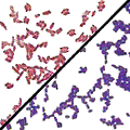
Gram staining is a method commonly used to determine the chemical make up of the cell wall of bacteria. The cell wall can stain either positive or negative, depending on its chemistry. If the bacteria stains positive it will retain a purple/blue color. If the bacteria stains negative, the bacteria will not retain the purple/blue color, but rather have a pinkish/red color.
Procedure:
1. Smear and heat fix a clean microscope slide with your bacterial culture.
2. Put 10 to 15 drops of Crystal Violet stain on your bacteria smear and leave on for one minute.
3. Rinse the Crystal Violet stain off with water (from the washing bottle).
4. Put the Grams' stain on the smear and leave it on for one minute, then rinse it off with water from the washing bottle.
5. Add 10 to 15 drops of 95% ethanol and leave on for 10 to 15 seconds. Rinse off with water for the washing bottle.
6. Place the Safranin on the slide and leave on for 45 seconds. Rinse off with water from the washing bottle. Add a cover slip.
7. Look under the microscope. Determine whether or not your bacterial culture stained positive or negative.
Questions
- Why is it important to follow sterile technique?
- What do gram-positive bacteria appear?
- Name three gram-positive bacteria and the diseases they cause.
- How do gram positive bacteria differ from gram negative ones?
In 1883-4 Dr. H. C. GRAM, a physician who was working with R. Koch, discovered that bacteria fell into two distinct categories when stained sequentially with CRYSTAL VIOLET followed in sequence by a bath in an IODINE SOLUTION, a wash with a destaining agent and a COUNTER-STAIN, if the cells were bathed following an initial treatment with the crystal violet. One group of cells RESISTED the removal of the crystal violet when washed with ETHANOL or ACETONE (DECOLORIZING AGENTS), whereas the second group was readily DECOLORIZED by a brief rinse with these reagents. To visualize the decolorized cells Gram briefly exposed them to a COUNTER-STAIN, or a stain of a different color from the crystal violet. Gram settled on the red counter staining dye SAFRANIN. Thus cells which resisted decolorization remained DEEP PURPLE or BLUE and came to be referred to a GRAM POSITIVE cells, whereas cells that easily lost the crystal violet dye were red after counter staining. These red cells came to be called GRAM NEGATIVE cells.
Gram and others drew several important conclusions from these staining results. First, they recognized that this staining was DIAGNOSTIC and could be used to IDENTIFY CELLS and SUBSTANCES. Secondly, they reasoned that cells and cell components DIFFERED CHEMICALLY as evidenced by their differential staining. Ultimately these realizations led to the idea of a MAGIC BULLET, which eventually led Fleming to recognize the significance of penicillin, which finally brought the world antibiotics and CHEMOTHERAPEUTIC AGENTS to treat diseases.
|
|
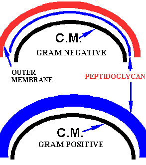
We now know that G+ BACTERIAL CELLS are very different from G- CELLS. By knowing this characteristic we automatically know a lot about a given bacterium; it is very much like knowing the "sex" of an organism because that one "fact" tells you so much.
Performing a good gram stain is easy, but it does require some experience. For example, the original "bacterial smear" must not be TOO THICK or TOO THIN or your results will be poor. Also the age of the culture is important. Very young and very old cells often produce poor results, whereas mid-log cells that are healthy and growing optimally, tend to give dependable results.
Manipulative Skills IB Biology Assessment Tick Sheet.
| Assessed Experiment Staining Bacterial Smears Manipulative |
washing hands and swabbing bench | sterile inoculating loop technique | 2nd Slide sterile innocc. | 3rd Slide sterile innocc. | 4th Slide sterile innocc. | Following the Gram Staining Method carefully | Focusing Microscope on x400 | focusing with oil immersion | |
