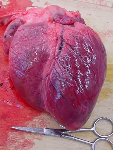
 Ventral side of sheep's heart |
Preliminary Discussion Questions 1) What is the heart's surface like? Why does it stop the heart
from
|
Heart Dissection Name Date
Aim
To study the anatomy of the heart and to relate the structures to their
functions.
To assess IGCSE skill C1 (Experimental techniques and use of
apparatus) and C2 (observing, measuring, recording).
 Ventral side of sheep's heart |
Preliminary Discussion Questions 1) What is the heart's surface like? Why does it stop the heart
from
|
Method
You will be given a sheep's heart, a dissection board, dissecting tools.
First, work out which is the dorsal (back) and ventral (front) side of the heart. The ventral side is the most convex (rounded). The thick walled arteries come from this side too.
Look for the trachea and the oesophagus. - Before you remove them from the heart - draw a cross-section diagram of each on a piece of A4 paper.
| Oesophagus.
Notice the thick layer of muscle used for peristalsys. |
 |
Trachea.
Notice the cartilage thickening to stop the trachea from collapsing like a whoopy cushion. |
Identify the parts of the heart shown in the diagram below: The side shown in this diagram is the ventral side (rounded side). Don't cut it yet.
|
Aorta |
 |
Pulmonary artery |
| The two vena cava go into the right atrium on the other side (dorsal side) | The pulmonary vein goes into the left atrium on the dorsal side. | |
| Coronary artery and vein | ||
| When you need to see inside the right ventricle, cut here. | ||
| When you want to open the left ventricle cut here. |
Identify the arteries and veins which come out of the heart. Measure
the diameter of the arteries and the thickness of the walls. Describe what
the arteries look like? (colour, texture, etc)
______________________________________________
_______________________________________________
_______________________________________________
Carefully put a rubber tube into part of the vena cave, close off any other holes in the vena cava and gently turn on the tap. From which blood vessel does water come out of the heart? This is the pulmonary artery
Repeat the last step with the pulmonary vein. The water should flow out of the Aorta.
Look carefully at the surface of the heart. Describe the pericardium
membrane ? Why is it shiny and slippery?
______________________________________________
_______________________________________________
_______________________________________________
Can you find any coronary arteries or veins. Where are they
found? What is their function?
______________________________________________
_______________________________________________
Cut open the left ventricle following the lines on the diagram.
Turn the heart upside down and run water into the ventricle. Can you see
the flaps of the bicuspid valve? Draw a sketch of one valve with the
suspensory ligaments on the A4 paper.
Cut the aorta leaving about 3cm above the heart. Run water into the
Aorta from the top of the heart. What structure can you see inside which
might stop the water? Why are they called "semi-lunar valves"?
______________________________________________
_______________________________________________
_______________________________________________
Cut open the right ventricle by following the lines on the diagram.
Describe how this is different from the left ventricle in term of volume and
muscle thickness. Measure the thickness of the muscle.
______________________________________________
_______________________________________________
_______________________________________________
Cut into the atria and measure the muscle thickness. Is the muscle wall
thicker or thinner than the ventricles.? Explain why this is the
case.
______________________________________________
_______________________________________________
_______________________________________________
Finally, what can you say about the size (volume) of each of the
chambers? Are they different sizes, which is the largest?
______________________________________________
_______________________________________________
_______________________________________________